Tin tức
WHAT ARE THE DIFFERENT TYPES OF DENTAL X-RAYS?
Currently, There are three types of diagnostic radiographs taken in today’s dental offices:
- Periapical X-rays ( also known as intraoral X-rays or wall-mounted X-rays)
- Panoramic X-rays
- Cephalometric X-rays
What are the different types of dental X-rays? We can take a look at some points below.
- Periapical X-rays
Periapical X-rays are probably the most popular, with images of a few teeth at a time captured on a small film cardinserted in the mouth in order to identify tooth status in the dental treatment. (They can be Portable X-rays or Wall-mounted X-rays).
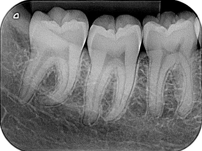
Image 1: Periapical X-rays image
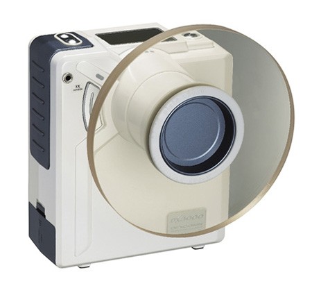
Image 2: Portable X-rays DX3000
2. Panoramic X-rays/ Pan
This is a wrap-around radiographic image of the patient’s mouth. The general film size is 5” x 11” ( or 15cm x 30 cm). Panoramic X-rays very useful for studying the patient’s jaw and the position of the teeth relative to one another. As previously mentioned, there are many additional regions of patient’s anatomy that can be imaged with a panoramic machine. (tissue /sinus). This machine is placed in the dental x-ray room (dental lead room).
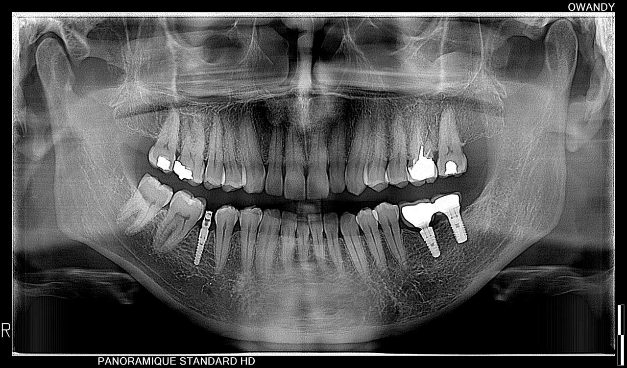
Image 3: Panoramic X-rays image
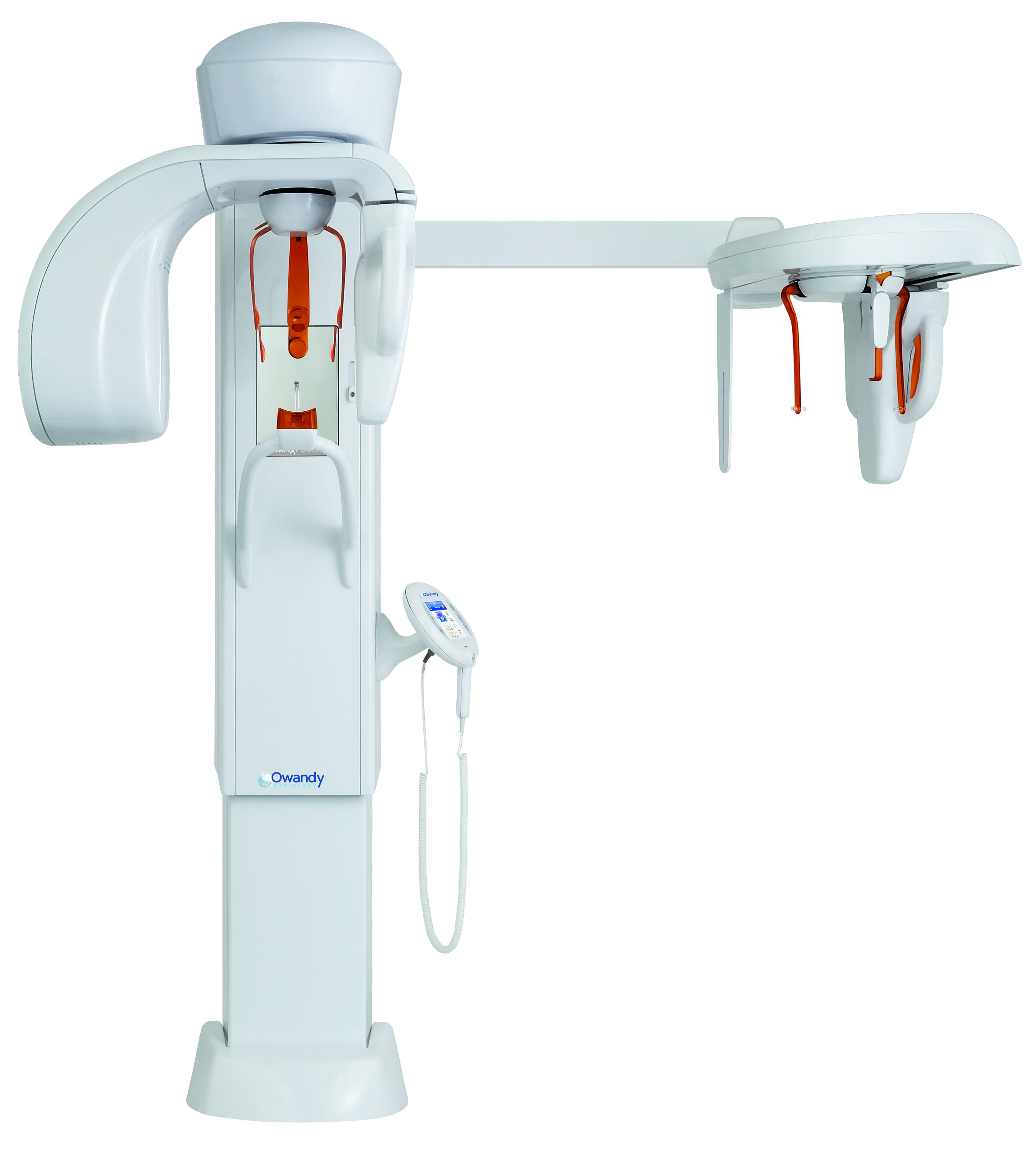
Hình 4: Panoramic X-rays machine
3. Cephalometric X-rays/Ceph
X-rays capture a radiographic image of the entire head, usually in profile. These films are most often used by orthodontists to diagnose misalignment of the jaw and bite problems. Ceph images are taken on a standard panoramic machine outfitted with a cephalometric film-holding arm mounted off to one side.
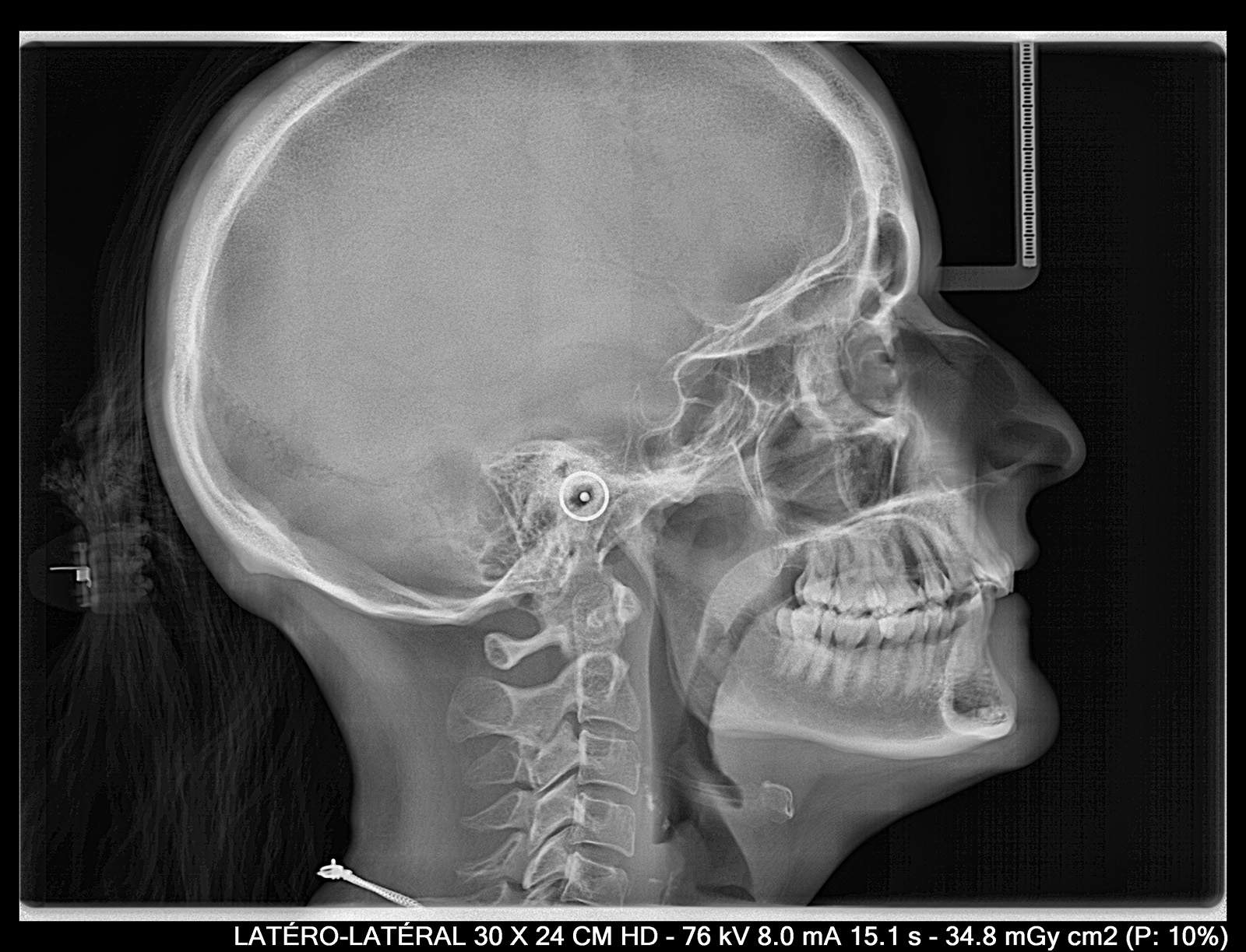
Image 5: Ceph image
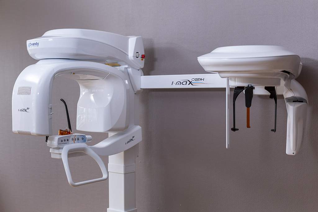
Image 6: Ceph machine


